|
Julia thecaphora (Carpenter, 1857) |
|||
|
¡¿A bivalved gastropod from Mexico?! |
|||
|
|
|||
|
|
|||
|
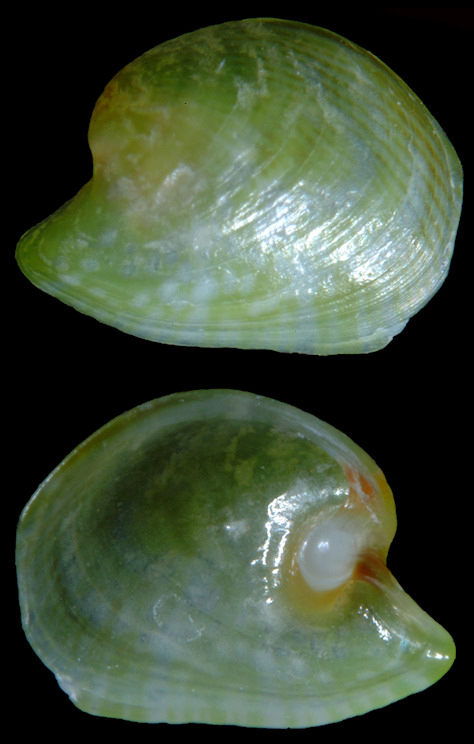
San Agustinillo, Oaxaca, Mexico. Both specimens depicted above are views of the interior and exterior of a single valve and measure about 3 mm. Digital images by David Kirsh. |
|||
|
|
|||
|
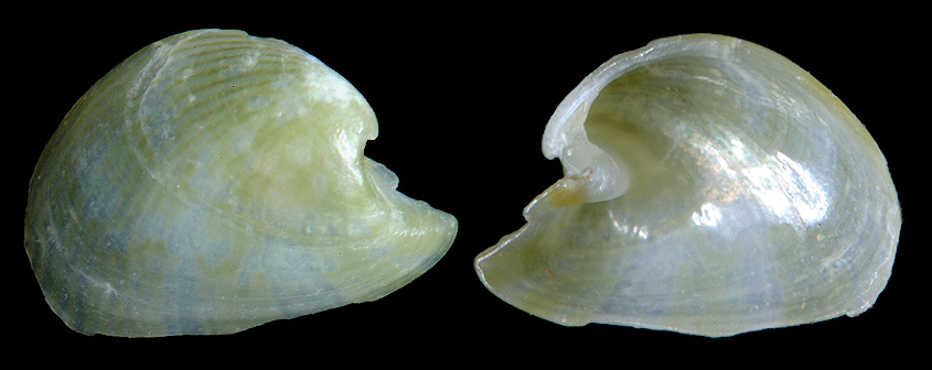
San Agustinillo, Oaxaca, Mexico. Digital image by David Kirsh. |
|||
|
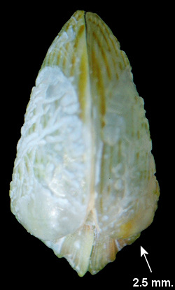
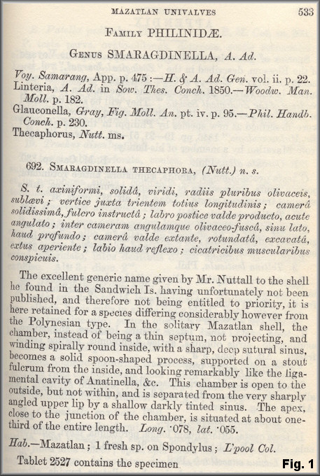
A note about the nomenclatorial and taxonomic history of Julia thecophora
This species was regarded as a pelecypod for most of the century
following its description. However, the solitary specimen Rev.
Philip Carpenter had before him, a right valve, was assigned to the
genus Smaragdinella A. Adams and Reeve, 1848 [TS Bulla
viridis Quoy and Gaimard, 1832 (= Bulla calyculata
Broderip and G.B. Sowerby I, 1829)] as evidenced in the original
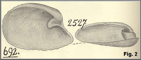
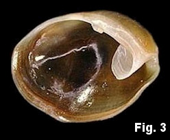
description (Carpenter, 1857: 533) [Fig. 1]
and its drawing by the author [Fig. 2 ].¹
The hinge
process of this specimen, which Carpenter likened to as a "theca"
[Greek theikos, meaning sheath], is apparently homologous to the "calycula"
[diminutive of Greek kalyx, little cup] of the univalve shells of
that genus as exemplified by the type species [Fig. 3], and recapitulated in
its specific epithet.
¹All sixty plates of these drawings languished in the
archives of the U.S. National Museum for over a century until being
organized and published (Brann, 1966), from which Fig. 2 is taken. |
|||